What it is
CL is caused by a bacterium called Corynebacterium pseudotuberculosis. It enters the body through breaks in the skin, such as scratches from thorny fences, ear-tagging, castration, dehorning, shearing or clipping wounds, and fighting. It can also enter through the mouth or nose. The bacteria travel to the nearest lymph node. There they hide inside white blood cells and are protected from the body's defences. The body walls off the infection with a thick capsule, and a slowly growing abscess forms. It is filled with pus that becomes thick and cheesy, which is what 'caseous' means. In goats the pus is often pasty and cream to greenish in colour. When an abscess bursts, the pus carries huge numbers of bacteria onto the skin, the ground, feeders and walls. In some goats abscesses also form in internal lymph nodes and organs, mostly the lungs. These cannot be seen from outside. They slowly cause weight loss, coughing and poor production. This hidden form is sometimes called 'thin goat' or 'wasting' disease.
Which animals are most at risk
Any goat can be infected, but signs are most often seen in adult goats over about one year old. The abscesses take weeks to months to grow, so infections caught when young often show up later. Herds that buy in animals from markets or other farms without checking them are at higher risk. So are herds that share bucks or use communal grazing and watering points. Dairy goats in zero-grazing units are at risk because they are housed close together and share feeders. Commercial meat herds that are handled often for tagging and castration are at risk too. Thorny bush grazing in pastoral and semi-arid areas leads to many skin scratches that let the bacteria in. Sheep kept with goats can carry and spread the same infection. There is no strong seasonal pattern. Spread can rise when animals are crowded, such as in dry-season holding pens or wet-season indoor housing, and after handling events like castration, dehorning or shearing of sheep.
Signs to look for
Early signs
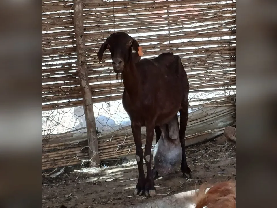
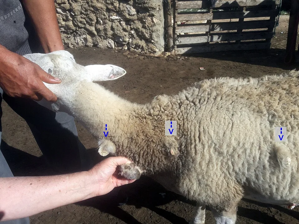
- A single firm, round lump appears under the skin at a lymph node site. Common places are below the ear, under the jaw, at the angle of the jaw, in front of the shoulder, in the flank in front of the hind leg, above the udder or near the scrotum.
- The lump grows slowly over weeks and is usually not very painful. The goat often keeps eating and behaving normally.
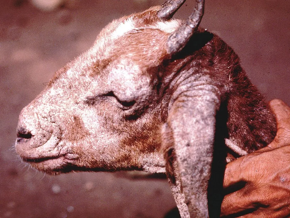
- The skin over the lump may start to thin and lose hair. The centre may feel softer than the edges as the pus matures.
- More than one lump may appear on the same goat, or similar lumps show up on several goats in the herd over months.
- Some goats show no outside lumps at all but slowly lose condition despite good feeding. This can be an early sign of internal abscesses.
As it gets worse
- The abscess bursts and leaks thick, cheesy, cream to greenish pus with little smell. This leaves a wet, crusted, hairless patch.
- Burst abscesses often heal over and then come back at the same site or at another lymph node months later.
- Large abscesses near the throat can press on the windpipe or gullet. This causes noisy breathing or difficulty swallowing.
- The goat loses weight steadily, has a dull coat and a lower milk yield, even while still eating. This happens when abscesses form in the lungs, liver, kidneys or internal lymph nodes.
- A chronic cough, faster breathing or tiring easily can point to abscesses in the lungs.
- An abscess in the udder area can lower milk yield and may affect milk quality.
- In severe internal cases the goat becomes very thin and weak and may eventually die. This is slow, usually over many months.
What to do right now
- Separate any goat with a lump or a burst abscess from the rest of the herd straight away. Keep it in a pen that can be cleaned easily, away from shared feeders and water points.
- Do not squeeze, cut or burst the abscess yourself. This spreads millions of bacteria onto the ground and your hands, and spoils the sample the vet needs to confirm the disease.
- If an abscess has already burst, wear gloves. Wipe away the pus with paper or cloth, then burn or bury the waste deeply. Keep the goat isolated until the wound has fully healed and dried.
- Clean and disinfect the area where the goat stood, as well as any feeder, wall or post it rubbed against. Remove the bedding and burn it if you can, and wash your hands and boots afterwards.
- Check every goat in the herd by running your hands over the jaw, neck, shoulders, flanks and udder or scrotum. Write down each goat with a lump, the site and the date.
- Stop selling, buying, lending or sharing animals, especially breeding bucks, until your vet has advised you. This avoids spreading CL to other farms or bringing more in.
- Make sure the isolated goat has clean water, good feed, shade and shelter so it keeps its condition while you wait for the vet.
- Call your vet or County Veterinary Officer to examine the goat, take a pus sample and plan how to handle the abscess and protect the rest of the herd.
How it spreads
- Pus from a burst abscess is the main source of infection. It contaminates bedding, soil, feed troughs, water points, fences and walls where goats rub.
- The bacteria can survive in the environment for months, especially in shaded, damp or dusty bedding and on wooden structures. A contaminated pen stays risky long after the sick goat has gone.
- Goats pick up infection through small wounds. Common ones are thorn and wire scratches, ear-tag and tattoo holes, castration and dehorning wounds, and injection sites made with dirty needles.
- Shared equipment spreads it from goat to goat. This includes clippers, shears, hoof knives, needles and tagging pliers that are not cleaned and disinfected between animals.
- Head-butting and rubbing against each other, feeders and posts spread pus between goats, especially in crowded pens and at feeding time.
- Goats with abscesses in the lungs can cough out bacteria. Nearby goats in close housing can then breathe them in.
- Buying an infected goat, often one that looks healthy because the abscess has not yet appeared, is the commonest way CL first arrives on a farm. Shows, markets and borrowed breeding bucks are also high-risk.
Could it be something else?
Abscesses from other bacteria or dirty injections
Many bacteria can cause lumps, especially at injection sites on the neck or thigh. These usually appear soon after an injection or wound, not at a lymph node, and often have smelly, runnier pus. Morel's disease, caused by a Staphylococcus germ, looks very similar to CL. Only a lab culture of the pus can tell them apart for certain.
Bottle jaw from worms (haemonchosis)
Bottle jaw is a soft, fluid-filled, doughy swelling under the jaw that often shrinks during the day when the goat grazes head-down. The goat also has pale eyelids and gums from blood loss to barber's pole worm. A CL abscess is a firm, defined lump at a lymph node, and the eyelids are normally pink.
Goitre or salivary cysts
Goitre is an enlarged thyroid gland caused by iodine deficiency. It shows as a smooth swelling on both sides of the upper throat, most often in young kids, and does not burst or produce pus. Salivary cysts are soft and fluid-filled. A vet can tell them apart with a needle sample.
Tuberculosis, tumours or other causes of wasting
Goats with internal CL lose weight much like goats with TB, Johne's disease, heavy worm burdens or tumours such as lymphoma. Outside abscesses elsewhere in the herd point towards CL. Faecal tests, blood tests, worm egg counts or post-mortem by your vet are needed to be sure.
How it is diagnosed
A vet usually suspects CL from the typical firm lumps at lymph node sites and the thick cheesy pus. To confirm it, the vet takes a sample of pus with a sterile needle from an abscess that has not yet burst. The sample goes to a laboratory, such as a County or regional veterinary lab or a KALRO or university lab, to culture and identify Corynebacterium pseudotuberculosis. This matters because other bacteria can produce similar abscesses. Some laboratories offer blood tests, such as ELISA, that detect antibodies to CL. These help find infected goats that have no visible lumps and are useful when screening a herd or animals you plan to buy. The blood tests are not perfect, and availability in Kenya varies. Internal abscesses can sometimes be seen by ultrasound or X-ray, but often they are only found at post-mortem or at the slaughter slab. Before the vet visits, write down which goats have lumps, where the lumps are, when you first noticed them and whether any have burst. Also note any goats that are losing weight. Keep the affected goat penned separately, and do not burst or squeeze the abscess, because a closed abscess gives the best lab sample. Have clean water, a secure place to hold the goat and records of recent purchases, tagging, castration and treatments ready.
Treatment
There is no quick cure for CL. The bacteria hide inside a thick capsule where medicines reach poorly. Treatment aims to reduce suffering, limit spread and help you decide which goats to keep. For a mature outside abscess, the vet may lance and drain it in an isolated area away from the herd. The vet collects and destroys all the pus and flushes the cavity with an antiseptic, often an iodine-based solution. The goat then stays isolated until the wound has healed completely, which can take a few weeks. Sometimes the vet removes the whole lymph node surgically under local anaesthetic, or uses other techniques to manage the abscess. Antibiotics, for example penicillin-type drugs, macrolides or tetracyclines, are sometimes used alongside drainage, especially for valuable animals. They often do not clear the infection on their own, and abscesses commonly come back. Responsible antibiotic use matters: using antibiotics that are unlikely to work, or not completing the course your vet prescribes, adds to antimicrobial resistance and wastes money. Any medicine must be prescribed by a vet, who will decide the drug, dose and length of treatment. All treatments carry milk and meat withdrawal periods. These are the time after treatment during which milk must be thrown away and the animal must not be slaughtered. Ask your vet for the exact periods and follow them. For goats with repeated abscesses, internal abscesses or severe weight loss, the vet will often advise culling, because these animals keep infecting others and rarely recover fully. Your vet can also help you plan a herd control programme. This may include blood testing, culling positive animals, cleaning and, where a licensed vaccine is available, vaccination.
Medicines must be prescribed and dosed by a vet. Always respect milk and meat withdrawal periods, and finish the full course of any antibiotic.
Prevention
- Buy goats only from herds known to be free of CL. Check every new animal carefully for lumps, and ask for blood test results if available. Keep newcomers in a separate pen for at least a month and check them again before they join your herd.
- Check all goats regularly for lumps at the jaw, neck, shoulder, flank, udder and scrotum, for example once a month and at every handling. Isolate any goat with a lump at once, before it bursts.
- Handle abscesses only under vet guidance, away from the herd. Collect and burn or deeply bury all pus, dressings and bedding from affected goats.
- Cull goats that have repeated abscesses or are wasting from suspected internal CL. Do not keep or sell them as breeding stock, because they keep the disease going.
- Reduce skin wounds. Remove or cover nails, sharp wire and broken boards in pens, and trim thorny branches near paths and feeding areas. Avoid overcrowding, which leads to fighting and butting.
- Use clean, disinfected equipment for ear-tagging, castration, dehorning, hoof trimming and clipping. Use a new or sterilised needle for each animal, and handle the youngest and healthy goats first and any suspect goats last.
- Clean and disinfect pens, feeders, water troughs and rubbing posts regularly. Allow sunlight into goat houses, because sunlight and drying help reduce bacteria in the environment.
- Ask your vet whether a CL vaccine is available and licensed for goats in Kenya, because availability is limited and changes over time. Where vaccines are used, they reduce new cases but do not cure goats already infected.
- Keep good records of purchases, lumps found, treatments and culling. These help you and your vet see whether control is working.
Risk to people
CL can occasionally infect people, though this is rare. It mainly affects people who handle pus or infected tissue, such as farmers, vets, butchers and slaughterhouse workers, and can cause swollen, painful lymph nodes that need medical treatment. Always wear gloves when touching a burst abscess or cleaning affected pens, cover any cuts on your hands, and wash well with soap afterwards. Keep children away from goats with burst abscesses. The bacteria can occasionally be found in milk from goats with abscesses near the udder, so always boil or pasteurise goat milk before drinking it. Meat from goats with abscesses should be inspected, and abscessed parts should be removed and not eaten. If you develop a painful swelling after handling infected goats, see a doctor and tell them you work with goats. Where antibiotics are used, follow the milk and meat withdrawal periods to avoid drug residues in food.
Impact on your farm
CL is a slow, money-losing disease rather than a sudden killer. Goats with internal abscesses lose weight and give less milk, which hurts dairy goat farmers in particular. Meat goats grow more slowly and fetch lower prices. At slaughter, abscessed carcasses or parts of carcasses may be trimmed or condemned by meat inspectors, and scarred skins lose value for the leather trade. Breeding stock from a herd known to have CL is harder to sell, and buyers increasingly check for lumps. Repeated vet visits, drainage, isolation and the loss of culled goats all add cost. Infected animals sold through markets carry the disease to other farms, which damages the seller's reputation and spreads losses across the community.
Outlook and recovery
For one goat with a single outside abscess, the outlook is fair. After proper drainage and isolation the wound usually heals within a few weeks, and the goat can stay productive. However, the goat remains infected, and new abscesses commonly appear later at the same or another site. Goats with many abscesses, abscesses that keep coming back or internal abscesses causing weight loss have a poor outlook and are usually best culled. At herd level, CL tends to stay once it is established, because the bacteria survive in the environment and in goats without visible lumps. With steady effort over several years, it can be brought under control and sometimes cleared. That effort means regular checking, isolating and culling, strict hygiene, careful buying and vaccination where available. Success depends mostly on how consistent the farmer is.
Common questions
Can I just burst the lump myself and squeeze out the pus?
Please don't. Bursting an abscess in the herd spreads huge numbers of bacteria onto the ground, feeders and your hands. Other goats can then be infected for months. It also destroys the closed abscess your vet needs for a lab sample. Isolate the goat and let the vet drain it away from the herd, with all the pus collected and destroyed.
Will antibiotics from the agrovet cure CL?
Usually not on their own. The bacteria sit inside a thick capsule where antibiotics reach poorly, so lumps often come back even after injections. Using antibiotics without a vet's advice wastes money, can leave residues in milk and meat, and adds to drug resistance. Your vet may use antibiotics alongside drainage in some cases, and will tell you the correct drug and withdrawal period.
Is it safe to eat meat or drink milk from a goat with CL?
Meat from a goat with CL should be inspected. Abscessed parts must be cut away and destroyed, and badly affected carcasses may be condemned. Never eat the pus or abscessed tissue. Always boil or pasteurise goat milk, and do not use milk from goats with abscesses on or near the udder until your vet advises. If the goat has been treated, wait until the full withdrawal period has passed.
Should I cull every goat that gets a lump?
Not always, but often it is the wisest choice. A goat with one abscess that heals well after vet drainage may be kept, isolated and watched closely, especially if it is valuable. Goats with repeat abscesses, several lumps or weight loss from internal abscesses keep infecting the herd and rarely recover, so culling them protects the rest. Discuss each case with your vet.
Can my sheep or cattle catch CL from my goats?
Sheep can easily catch the same infection from goats, and the two species often pass it back and forth on mixed farms. Cattle, camels and horses can be affected by related strains of the same bacterium, but they are less commonly part of goat outbreaks. On mixed farms, check your sheep for lumps too and apply the same hygiene and isolation rules.
Is there a vaccine for CL in Kenya?
Vaccines against CL exist in some countries, sometimes combined with clostridial vaccines, but they are not always available or licensed for goats in Kenya. Availability changes, so ask your vet or County Veterinary Officer what is currently approved. Even where a vaccine is used, it reduces new infections rather than curing infected goats. Buying clean stock, early isolation, hygiene and culling remain the backbone of control.
A guide, not a diagnosis. This page is general information to help you recognise problems early. Always confirm with a vet or animal health worker before treating.